


Welcome To
Flores Optometry Inc
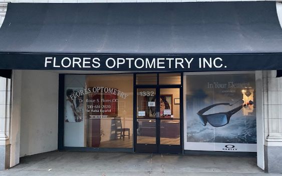
Flores Optometry Inc has proudly served the San Leandro community since 2007. And we want to help you achieve and maintain clear vision for years to come.
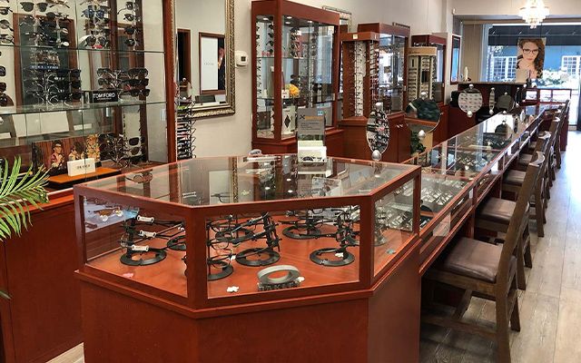
Our experienced team of eye care professionals offers comprehensive eye exams, quality lenses and frames, and more. By leveraging advanced diagnostic technology, we are committed to improving the quality of life of our San Leandro community through enhanced vision.
Give yourself the gift of clear vision — schedule your appointment with Flores Optometry Inc today.

Meet Our Doctors
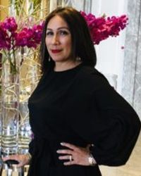
Dr. Rosario Flores, O.D.
Optometrist
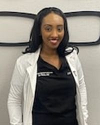
Dr. Lidia Tekie
Associate Optometrist

Dr. Helen Lam
Optometrist
State-of-the-Art
Our Technology


Eyewear Brands


Testimonials
Patient Reviews

Contact Info
Operating Hours
- Monday 9:00am - 5:30pm
- Tuesday 9:00am - 5:30pm
- Wednesday 9:00am - 5:30pm
- Thursday 9:00am - 5:30pm
- Friday 9:00am - 5:30pm
- Saturday 9:00am - 2:00pm*
- Sunday Closed
© 2024 Flores Optometry Inc. All rights Reserved. Accessibility Statement - Privacy Policy - Sitemap
Powered by:
Phron M.
Flores Optometry is the best optometry office I’ve ever visited. Dr. Tekie was not only professional, but she was knowledgeable, direct, and patient.
The Flores Optometry staff is very nice, professional, helpful, and care about their patients. They also have an impressive inventory of frames. Not only do they have great service, but their turnaround time on the production of glasses is impeccable.
I’m a fan. Thank you!!